
In this article
When you're working with lab-made protein fragments (called synthetic peptides) at your bench, you probably focus on the core sequence. That's the specific building blocks you need for your target. But new research suggests you're only seeing part of the picture.
A massive screening effort called ASHI (Atlas of SLiM-mediated Human protein-protein Interactions) has mapped over 20,000 interactions between human protein parts (called domains) and short protein pieces (peptides) from the disordered proteome. The study tested 800 protein domains against a library of about one million peptides covering the human disordered proteome. What they found challenges a common assumption: that short patterns (called short linear motifs or SLiMs) work all by themselves.
The Disordered Proteome Isn't Just Filler
Intrinsically disordered regions (IDRs) make up roughly a third of the human proteome. For years, these flexible, unstructured stretches were dismissed as biological noise, functionless padding that didn't warrant serious attention. That view has shifted dramatically.
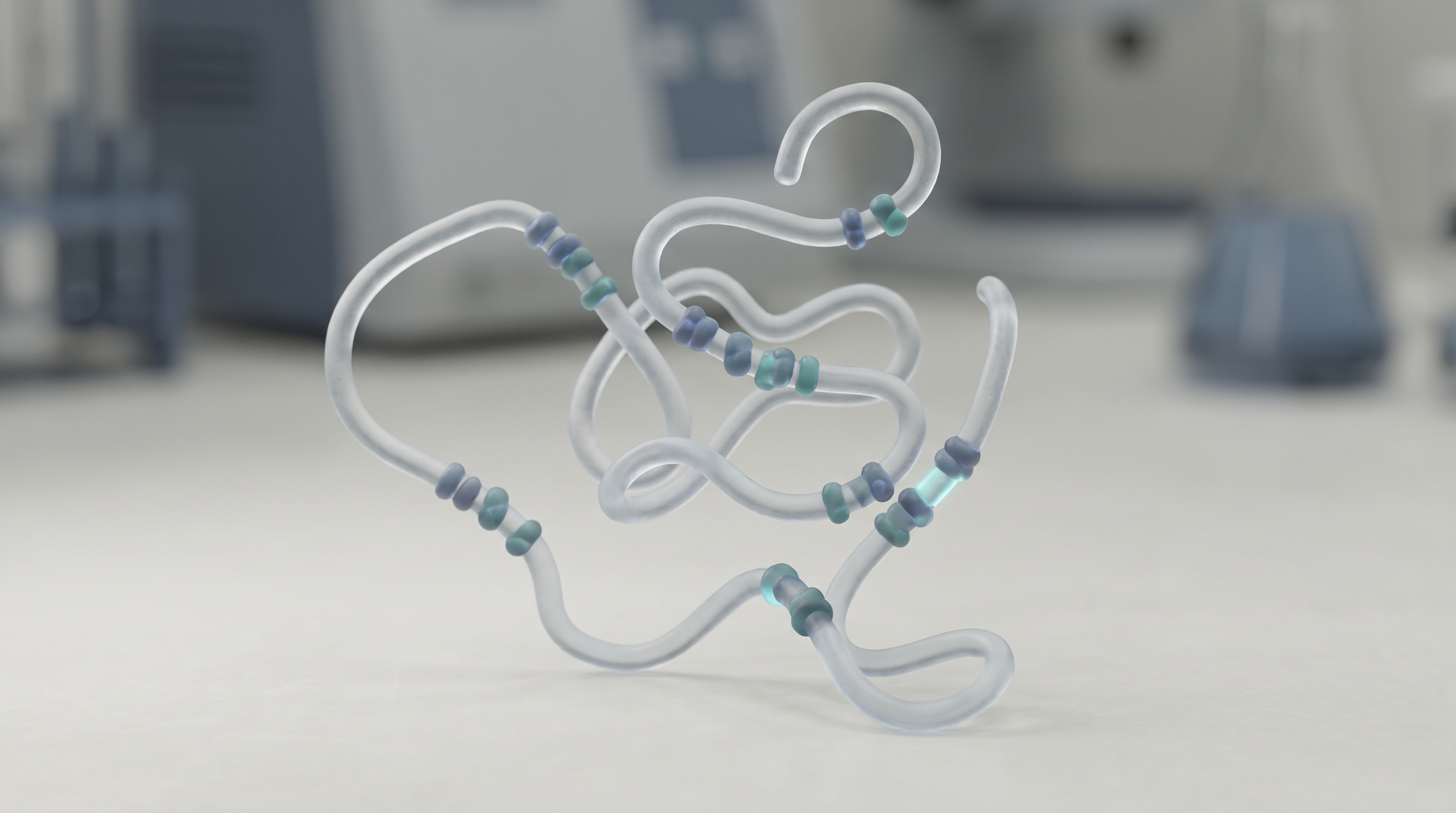
The ASHI data shows that IDRs work as densely encoded interaction platforms. A single disordered region can contain multiple overlapping short linear motifs. Each one can recruit different binding partners. But here's the finding that should catch your attention as someone working with peptides: interaction specificity is not determined by the motif alone.

Flanking Residues Are Key Determinants
The researchers found that residues (the individual building blocks) immediately next to the core short linear motif, positions you're probably not thinking about when you order your peptide, can dramatically change binding affinity. In some cases, changing a single flanking residue flipped an interaction from strong to undetectable.
Key point: Residues immediately next to a core short linear motif (SLiM) act as critical context-dependent determinants of binding affinity, rather than passive spacers.
This has practical implications. If you're reconstituting a peptide based on a motif you found in a database, the sequence context matters. The exact flanking residues you include (or leave out) in your synthetic peptide can determine whether you're faithfully recreating the native interaction or missing it entirely.
Expanding the Peptide-Binding Universe
The study also uncovered novel binding modes for known peptide-binding domains. Perhaps more striking was what they found in unexpected places: enzymes, chaperones, RNA-binding proteins, and modification-reader domains all showed peptide-binding activities that weren't previously documented. This suggests the peptide-binding universe is far larger than the textbook families you'd find in a typical binding domain database.
What This Means for Your Benchwork
If you're designing experiments around synthetic peptides, consider these takeaways:
- Order longer peptides than you think you need: Including flanking residues on either side of your core motif may be essential for reproducing authentic binding behavior. Truncating to the minimal functional sequence could eliminate context-dependent interactions.
- Match the native flanking context when possible: If your peptide corresponds to a natural protein region, try to preserve the native sequence surrounding the motif. The ASHI data shows this isn't optional, it's often the difference between a functional and non-functional interaction.
- Watch for off-target binding: The unexpected peptide-binding activities discovered in enzymes and other non-canonical binding proteins mean your peptide might interact with targets you didn't design for. If you're seeing unexpected results, consider whether flanking-region-mediated interactions could be at play.
The ASHI resource provides an unprecedented foundation for understanding these context-dependent interactions. It's freely available and maps interactions across the human SLiM interactome. For peptide researchers, it's becoming an essential reference for interpreting what you're actually seeing when your peptide meets its target.
Prompted by this coverage at bioRxiv →
Related from our lab: the pen · cartridges · bacteriostatic water · peptide calculator
Shared by Preppin Peppers for research, educational, and demonstration awareness only. We link to third-party coverage; we do not endorse it, and nothing here is medical advice or a recommendation to use any substance in humans or animals. Our products are sold for laboratory research use only.
Reminder: research and educational reference only. Preppin Peppers sells hardware and materials, not peptides. Not medical, dosing, or health advice, not evaluated by the FDA, and not intended for human or animal use.
Frequently asked questions
Why do flanking residues matter in synthetic peptide design?
Flanking residues adjacent to the core motif act as context-dependent determinants of binding affinity. Changing a single flanking residue can flip an interaction from strong to undetectable, making native context critical for reproducing authentic protein-peptide interactions.
What did the ASHI study reveal about peptide-protein interactions?
The ASHI study mapped over 20,000 interactions and found that interaction specificity is not determined by the motif alone. Residues immediately next to the core short linear motif dramatically influence binding affinity, challenging the assumption that motifs work independently.
How long should synthetic peptides be ordered for accurate binding studies?
Order longer peptides than you think you need. Including flanking residues on either side of your core motif may be essential for reproducing authentic binding behavior, as truncating to minimal functional sequence could eliminate context-dependent interactions.
More in our reusable reconstitution pens collection.
What the research community gets wrong about flanking residues and short linear motifs
Short linear motifs (SLiMs) are easy to look up in a database and easy to order as a peptide. That convenience hides a few common mistakes when you take a motif to the bench.
- A motif is not self-contained. The residues sitting right next to the core motif (the flanking residues) can change how the peptide binds in your assays. Studies report that context can contribute a meaningful share of the total binding energy, so treating the core as the whole story leaves interactions on the table.
- The shortest peptide is not always the correct peptide. Trimming a synthesized fragment down to the minimal core sequence can drop context-dependent interactions. A slightly longer fragment that keeps the native flanking region often behaves more like the real protein region in a binding measurement.
- A single-residue swap is not always harmless. Changing one flanking residue has been shown to move an interaction from strong to undetectable. If you order two nearly identical peptides and see very different results, the flank is a likely reason, not a failed pipetting step.
- Peptide binding is not limited to the textbook domain families. Screens have found peptide-binding activity in enzymes, chaperones, and RNA-binding proteins that were not on the usual list. An unexpected signal in your vial may be a real off-target interaction driven by the flanking sequence.
- Flanking sequence is not passive filler. These regions carry charged stretches and modification sites that tune both affinity and specificity. Leaving them out (or getting them wrong) changes what your peptide actually reports on.
From our bench: If you have run a core-only peptide next to a longer peptide that keeps the native flanking residues in the same binding assay, tell us what you saw. Report both sequences, your reconstitution buffer, and the direction of the difference (which one bound more, or whether the signal disappeared), with your actual numbers rather than estimates. Firsthand side-by-side observations from your vials help other researchers judge how much flank to include.
Sources
- Bacteriostatic Water for Injection, USP , FDA/DailyMed label (0.9% benzyl alcohol)
- Duerkop et al., Biotechnol J 2018 , Impact of Cavitation, High Shear Stress and Air/Liquid Interfaces on Protein Aggregation
- Sigma-Aldrich (Merck) , Handling and Storage Guidelines for Peptides and Proteins
- Palopoli et al., Short linear motif core and flanking regions modulate retinoblastoma protein binding affinity and specificity, Protein Engineering, Design & Selection 2018 (PubMed)
- ELM, the Eukaryotic Linear Motif resource, 2024 update, Nucleic Acids Research (PMC)
✔ Reviewed by Bryan Le, PharmD, RPh
Bryan is a licensed pharmacist (Doctor of Pharmacy, Registered Pharmacist). Reconstituting lyophilized preparations is core pharmacy practice, so he reviews The Lab’s content for technical accuracy and to keep it within a research-and-education scope, with no medical or dosing advice. View profile on LinkedIn.
